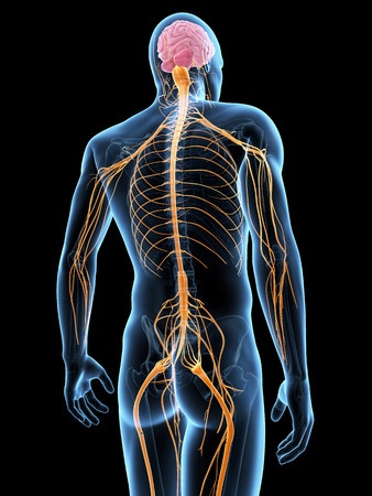
When I ask people in clinic to point out where their back is I get a variety of responses starting from the neck, down to the pelvis and anywhere in between. To most the spine is a blurry picture of a structure that sits some where between our head and hips. If it is not painful we often forget it is there. If it is painful we trawl the internet to find conflicting information about whether to flex it, extend it, brace it or just let our bellies hang out. Is pilates better than yoga? Should we swim or walk to ease it off? Never has there been so much confusing information available so readily at the end of a button. Let’s keep it simple and get to know your own back anatomy and how it all fits together
(P.S. latin words and anatomical names have been spared in the name of understanding all this stuff!)
The spinal column
The spinal column is made up of a series of bones (called vertebrae) all of which have a slightly different shapes and functions. These vertebrae all stack up to form the cervical spine (neck), the thoracic spine (mid back), the lumbar spine (lower back) and the coccyx and sacrum. A well functioning spine should move with fluidity and ease enabling us to walk, sit and dance (apart from my brother who could have a spine like a slinky and still dance like he has 3 left feet).
Each vertebrae is made of a dense bone outer shell with a lighter more honeycomb textured inner shell (malteser any one?) This structure allows our bones not only to be strong but also light, with ability to absorb force if we were to jump up and down or run. Each vertebrae is made up of a bony ‘body’ behind which you can find a hole called a foramen. When you stack all these vertebrae on top of each other the holes line up and become the spinal canal through which the spinal cord runs .
The spinal cord
The spinal cord ferries information from our peripheries to our brain and then returns information back from the brain telling all the various areas of the body how to react. The nerve ‘tributaries’ that carry this information are called spinal nerves and they wind their way back from all round our body and up through holes at the side of the vertebrae where they join the spinal cord. The information itself is detected by receptors that are attached to the spinal nerves and are scattered everywhere from the skin’s surface to inside blood vessels and organs. These receptors detect information such as temperature, pain, vibration, stretch and even tickle and itch. When you witness a dog pushing its side into you as you give it a good scratch you are seeing this relay of itch information to the brain which results in a messaged being sent back “go on muscles lean into it and enjoy yourself!”
Discs
In order to allow the spinal bones to move on each other and to absorb force, they are separated by a series of ‘rubber washers’ called discs. Each disc is made up of a fluid filled sac called a nucleus and a strong fibrous outer band called the annulus which covers it and glues it to the vertebrae above and below.
Interesting fact
These discs fill with water every night as we lie down making us a little taller when we first get up in the morning. This is why people with back pain are often advised against strenuous exercise or stretching first thing in the morning as the discs are already taught.
Disc ‘bulge’
When we have a disc ‘bulge’ the nucleus starts to protrude through a tear in the tough annulus coating and can eventually reach through into the spinal canal where it can press on the spinal nerves. When these nerves are pressed upon they create a predictable pattern of weakness, lack of sensation and pins and needles, very much like a hose which is kinked and unable to transport water. When this happens in the lower back it can press on the tributaries that make up the sciatic nerve, creating that familiar pattern of ‘sciatica’ that can refer all the way down into the feet.
Here is a nice little you tube animation which demonstrates what happens when you have a a disc issue.
https://youtu.be/33LsxW-Zq0s
Ligaments
Ligaments are like little straps that attach bones to other bones. The vertebrae are connected by capsules and ligaments which then help stabilise our bony spinal column. When these ligaments are continuously stretched (think bad sitting posture or a compensation around another injury such as an ankle sprain) then these ligaments can become painful and lax. When ligaments experience a gentle stretch they can feed back information telling the brain which muscles need to work, but over stretch them and they become unhappy. A lot of what we do in clinic is to work out why these ligaments keep getting stretched, then we use hands on techniques and movement to straighten everything up and allow the body to find its centre.
Muscles
Muscles attach all the way up the spine at various different depths and, very much like ligaments, need to experience being loaded as well being unloaded. If they are chronically stretched (for example if someone has their work desk set up so they are always rotated to one side) the stretched muscles will start to become painful and prone to tearing, while the shortened muscles will become congested. By finding a more balanced resting posture we can then remove the trigger to tissue pain and allow the body to do what it does best and start to heal.
Conclusion
So that was a quick whistle stop tour of getting to know your back anatomy. Once you know what is lurking around in that unknown area between your head and your pelvis, then things becomes a whole lot less scary. As a therapist all we do is find out the trigger that keeps causing things to be painful and remove it for a while while your body naturally gets better. Add a dash of movement to synchronise bones, muscles, ligaments and nerves to coexist in harmony and we have our selves a treatment plan.
I hope this cleared things up a little bit and allowed you to picture what is going on back there. If you know any one who might benefit from this little guided tour, please feel free to share.


